Words. Sentences. Paragraphs. What do they have in common? A beginning. When you write and consume language, you play with the idea of a beginning. You brain bubbles up with key words. What do I want to say? What is there to say? Who do I want to say something to? What should I not say?
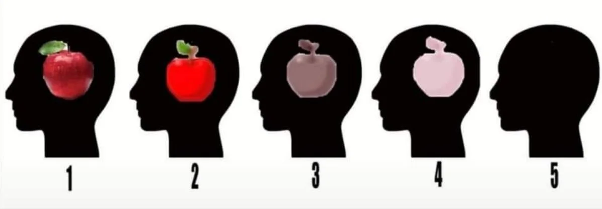
Each word begins to symbolize a neuron. Soon, you build a deeply connected linguistic neuronal network. Your temporal lobe springs to life. It becomes one big party where your broca’s area, caudate nucleus, and hippocampus are only a few of the invited guests. If you’re familiar with hosting gatherings, you know a good sense of order is key to avoid chaos. You can say the same about the brain and writing.
If you try to write, you can get overwhelmed. All of the parts in your brain activate and spit out a billion key words. A travel diary experience. The cool donut shop across the street. An elementary school teacher’s advice. The random wisdom you got on the subway. There’s plenty to write about. Your brain recognizes every story you can recall and brings them to your short term memory. Then what? You get confused because there are too many guests at this lexicon party. Hm! So the next logical step is to kick a few of them out and set some boundaries. They add to the chaos, instead of the story.
Turns out, filtering is the most important step when you write. It helps you choose a worthwhile beginning. Not only to yourself but to the reader on the other side. I learned this the hard way after I took a couple of writing classes. Too many ideas and you lose the focus of the message. Slowly, I learned how to uncover hidden gems in my linguistic neuronal networks. Stories I forgot about. Only to rediscover them after I learned how to filter and write the proper way (with rules — *gasp*).
To write is to organize your mind. To write, is to think. To think, is to live. In this article, I share a few tips my professional writing professors taught me to help create a fun linguistic brain party (I also got an A and got my writing published in a journal! along the way). It takes time to write better but the process can be rewarding. Let thy writeth commenceth!
Rule 1: Write in Drafts
If you want to never write anything, tell yourself you’ll write everything you want perfectly on the first try. That’s the recipe to guaranteed failure. Do you want to actually write something that can be good? Write in drafts. Try to aim for 4 drafts for any piece you want to write.
Draft #1 is when you brain dump. Write anything you can think of relative to the topic. Don’t think of anything else but writing it down. Don’t think about editing, grammar, references, thesaurus, facts, or anything else. Your sole objective is to dump everything on your current mind. You can set a 12 minute timer and get a pen and notebook to do this. You can’t erase words or wait to think of better words when you write in this stage. You write something and want to change your mind? Too bad, scratch it out and keep going. That’s the sole purpose of Draft #1. Get words down. Get out of your head and write anything, even if it’s “trash”. You can even use the notes app on your phone as a starting point. The job of the first draft is to just exist. Give yourself permission.
Draft #2 is when you edit the basics of what you wrote. Look at grammar, ways to paraphrase, and make your writing concise. Remove the unnecessary. Each sentence should hold an idea. Remove the elements that cloud that idea. Good writing is clear writing. Avoid “word salading”, word fluff, thesaurus alternatives, and distracting punctuation. Replace long sentences with short ones. Strive to have short sentences. Short is clear. Long is annoying and hard to comprehend and a waste of reading and confusing and unfocused in its messaging. See my point? Avoid adding more than one “and“ in a sentence. Turn it into two sentences instead. Have fun with editing in Draft #2.
Draft #3 is when you share your work with another person for feedback. This draft is critical because what makes sense to you may not translate to other people. Remember, when you write you try to have a conversation with another person. Make sure they can understand you. You do that by asking for feedback on your writing.
Draft #4 is when you strengthen your writing with external references. If you didn’t mention other research sources, you can add them here. Now that you know what you want to say, look at what else can be said in relevant fields. You can play with this draft a lot and introduce new perspectives or even collaborate with other authors. Make sure to assess the credibility of your sources and choose a reference style so a reader can access them if they’re interested in the details. Remember, your writing starts a conversation so give the reader opportunities to continue it elsewhere.
Four drafts are a good starting point. If you feel like four drafts are too high, then aim for “two drafts and a polish” as a minimum revision process. You can add more depending on how long you want to write for. Feel free to adjust the number of drafts ∝ quality and quantity of your messaging. Have fun with it and nurture your editing circles. You can encourage your friends to also write and host weekly editing circles to improve your writing skills together. It’s one of the best ways to forge strong friendships rooted in critical thinking and mutual respect.
Rule 2: Use Active Voice
A simple formula. A = S + V + O. Active Voice = Subject + Verb + Object. You want to write in active voice to be clear and confident with your messaging. Avoid Passive Voice. P = O + V + S. Passive Voice = Object + Verb + Subject. Don’t write in passive voice because you’ll often use dead verbs, gerunds instead of infinitives, and be unnecessarily wordy. Instead of passive voice, you want to write with prose — spoken language in its ordinary form.
That being said, “learn the rules like a pro, so you can break them like an artist“. For instance, you can use active and passive voice strategically if you write a story and want to show character personalities. A secretive character can speak with passive voice to be less direct and can rely on wordiness to confuse others. Whereas a investigative character would interrogate them with active voice as they ask clear and concise questions. Outside of novels, please use active voice to write. Especially if you write scientific papers, please don’t use unnecessarily complicated academic language.
Rule 3: Vary Sentence Lengths
Short sentences should be your “go-to“. Aim for 10 words maximum in a sentence. Remember each sentence expresses one idea. Don’t pack more in. Give the reading breathing room to digest each sentence comfortably.
Long sentences can be used for “journey“ scenes. The longer the motion, the longer the sentence. If you write a long sentence, present the information in chronological order. You can also use parallelism to show how two or more elements in a sentence have the same level of importance. Parallelism only tends to work if the grammatical elements are the same and fit the story. What journey is the sentence going to try to express? Imagery of a vivid environment? A physical description of a character? An emotional monologue? If you use long sentences, be intentional about it. Place them in variation with other sentences.
Mix long and short sentences to shape your reader’s flow of information. For instance, use short sentences to slam the breaks on a story. It’s one of the best ways to add raw emotional power to a story. We all know those short one-liners that punch tears out of our eyes. Use short sentences to deliver those when needed. You can also use long-to-short sentences to add tension and release it in a piece of writing. You usually see these when you want to “conclude“ a moment or message at a certain section of your writing. To do the opposite, use short-to-long sentences to “introduce“ a moment. For example, you can expand and explain a certain topic. Or you can draw out a feeling in your readers with a sharp to ambient moment expressed with imagery. It’s all about the message and feeling you want to create.
You can think of sentence length as a frame in a movie. How many things do you want to show in a frame and for how long? What moments do you want the audience to absorb from the movie and which scenes can you afford them missing? It’s all about how you manage the reader’s attention. Luckily, sentence length helps you decide that.
Rule 4: Know When to Backoff
When you write, you need to balance when to back off versus show off. You can use understatements to let the reader pay attention to what’s happening in the story at the moment. You would want to use shorter sentences and dead verbs in this case. These intentional understatements metaphorically pass a “moment“ for your reader to think for themselves. You intentionally don’t give them much to work with. The reader needs to have room to enjoy and contribute to the story with their own imagination or emotions. The more serious or intense the subject is, the more you should back off in how you deliver the material.
Overstatements are the opposite. You let the reader pay attention to details you want to deliver vividly. You would want to use active verbs, parallelism, and the occasional freight train. The more playful or inconsequential the topic, the more you can show off an overstatement. Don’t use them in intense moments. Instead, use them to elaborate story details with wonderful language you can afford to play with like sentence length, rhythm, and thematic variety so the reader doesn’t get bored. Overstatements are good to include in between critical pieces of a plot while you build up to them.
To create a rhythm in your writing, balance understatements and overstatements. In a sense, aim to “deliver hot emotional content with cool hands“. Let your writing deliver its message and give enough breathing room for the reader to make their own conclusions. It’s a conversation after all.
Rule 5: Embrace Storytelling
Writing doesn’t have to be boringly serious all the time. Have fun with it! Never tell us a thing if you can show us instead. “Show, don’t tell” is the golden rule for superb writing. In the iconic “Writing Tools“ book, Roy Peter Clark says a good description is made of a few well-chosen details that will stand for everything else. For that, use cinematic paraphrasing. It’s when you read something and it reads like a movie. That’s the fun kind of reading!
Think of your notebook as a camera when you write your stories. Try to screenwrite. Use the standard camera angles like aerial views, an establishing shot, middle distance, close up, and extreme close up when you describe story events. Each camera angle has a different purpose and you want to create a new paragraph for each camera angle change. In a dialogue, each person changes the camera angle. If you want to choose a camera angle, think of this. Where is my narrator in the scene? Are they an appropriate distance away? A good rule of thumb is to have a diversity of camera angles in your writing.
An aerial view is where the narrator looks down on the world like being in a hot air balloon looking down. You can describe a scene from above with it. An establishing shot is when you stand back to capture a setting where action is going to take place in a story. You describe the world you’re about to enter, create a mood. A middle distance is when the camera moves closer to the action with key players and their interactions. It’s also the most common shot. A close up shot gets in the face of a character so the narrator can detect their emotional state. An extreme close up zooms in on a detail that is normally invisible from a further distance.
Rule 6: Incorporate Symbolism
Humans are meaning-making machines. We love to see beyond the physical world and stamp personality. Symbolism helps make that literal when you write. Creating your own symbols has a useful purpose as a focusing device. A reader should then know it instinctively after finishing your story. It’s a powerful tool when used correctly. Especially since a good idea tends to stick around, and stick around and stick around. That being said, avoid clichés. They’ve already stuck around into the collective psyche and don’t need to be reinforced in lieu of your fresh, new ideas or symbols.
Rule 7: Balance Input-Output
The two musts of any writer is to read a lot and write a lot. Balance your input with output. Be a voracious reader and you’ll naturally become a hungry writer. There’s a beauty to stories. You start to recognize them everywhere. Any moment can become a story. Especially since most stories are about a person who tries to get what they can’t have. That seems like one of the axioms of life, no? Channel that. Mind wandering can also be a time and place, if you make it so. Writing does that with refined thinking. While you look to create your own stories, read other people’s stories too. There are endless masterpieces to read across human history. Give yourself the opportunity to bask in that neural richness. It turns out, humans are pretty good at this storytelling thing. Perhaps, even the best. There’s no reason why you can’t join in on the fun too.
Just give yourself a chance. Trust me, improving your writing skills is the most empowering thing you can do in your life. It’s a shareable psychoscope. It helps you think clearer, share your internal world with others, build relationships, and everything else products usually try to sell to you on billboards. Pen and paper. It’s that simple. Have a conversation with yourself and see what beauty you can come up with. You’d be surprised. We all look for beauty in the world. Writing shows how a lot of it can come from you. The brain is a beautiful machine, give it a chance to show you. Write.